
Robotic Mediastinal Cancer Surgery in Ahmedabad
Dr. Nitin Singhal offers robotic-assisted surgery for mediastinal tumours in Ahmedabad, including thymectomy and mediastinal mass excision, bringing 18+ years of experience to precise, minimally invasive tumour removal.
Dr. Nitin Singhal
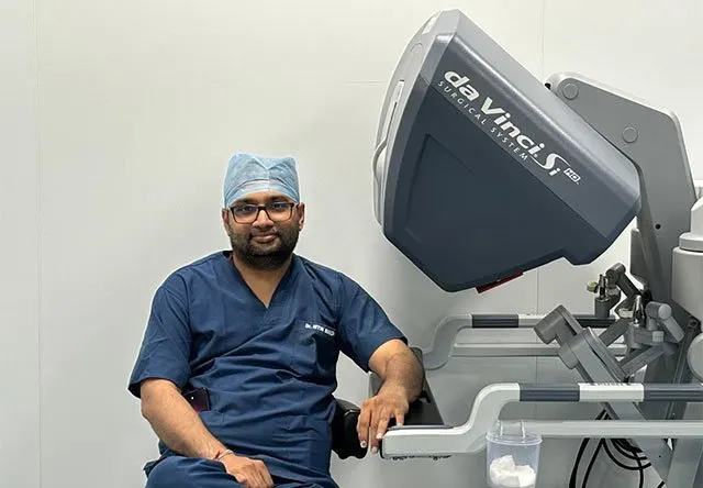
Dr. Nitin Singhal is a Robotic and HIPEC surgeon based in Ahmedabad, specialising in thoracic oncology, including complex mediastinal tumour surgery. He combines robotic-assisted technique with a patient-first approach, aiming for precise cancer removal alongside faster recovery wherever suitable.
What Is Mediastinal Cancer?
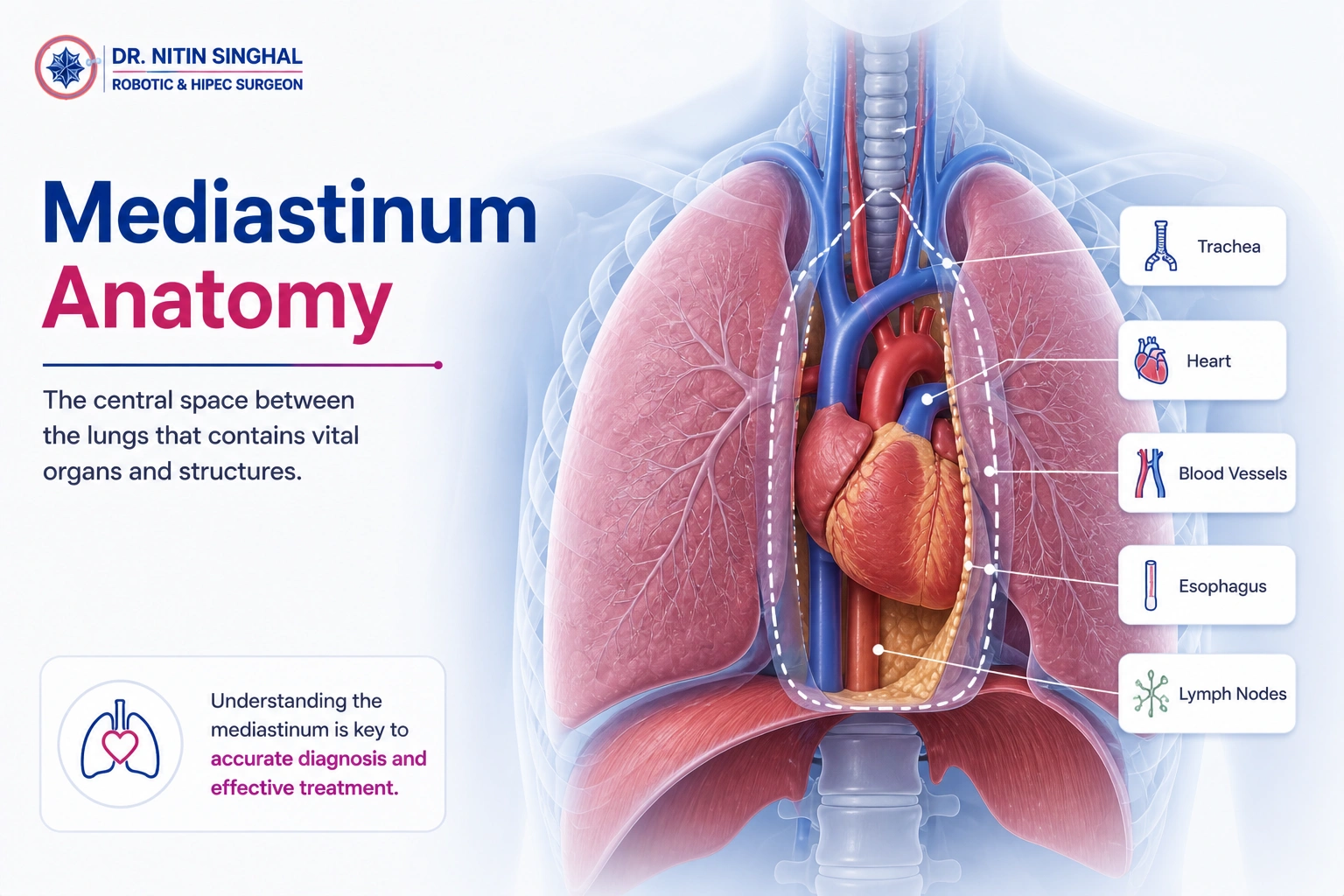
Mediastinal cancer refers to tumours that develop in the mediastinum — the central compartment of the chest that sits between the two lungs, housing the heart, major blood vessels, the thymus gland, part of the oesophagus, and the trachea. Tumours here are grouped by which part of the mediastinum they arise in — anterior, middle or posterior.
The most common types include thymomas and other thymic tumours, lymphomas, germ cell tumours, and neurogenic tumours arising from nerve tissue. Because the mediastinum sits close to the heart and great vessels, tumour location has a major influence on symptoms, diagnostic approach and the surgical technique needed.
Symptoms to Watch For
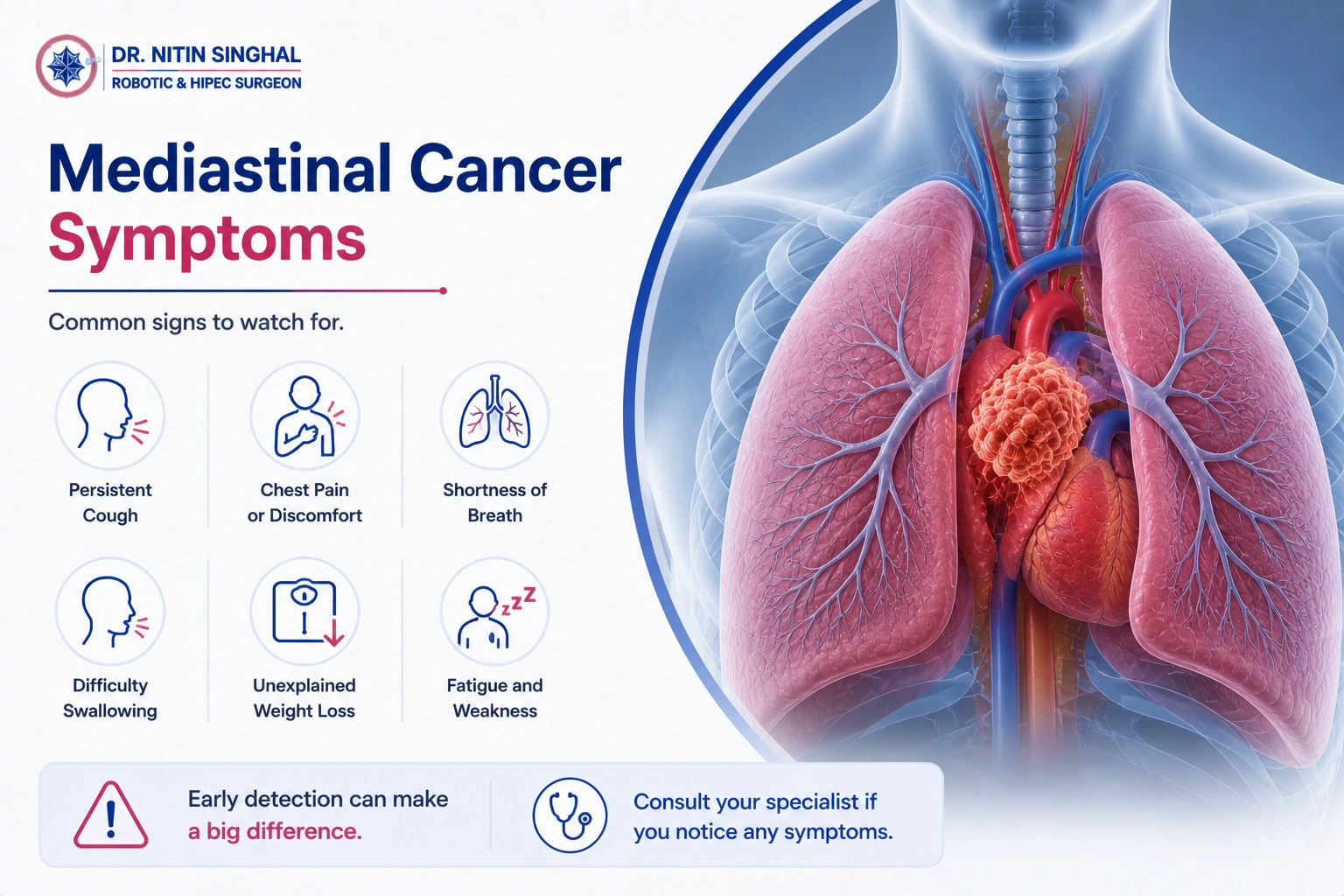
Many mediastinal tumours are found incidentally on a chest X-ray or CT scan done for another reason. When symptoms do occur, they often relate to pressure on nearby structures.
Chest Pain or Pressure
A dull ache or feeling of pressure in the chest, sometimes worsening with breathing.
Persistent Cough
A cough that doesn't resolve, sometimes with breathlessness.
Shortness of Breath
Difficulty breathing, particularly if the tumour is pressing on the airway.
Hoarseness or Swallowing Difficulty
Changes in voice or trouble swallowing from nerve or oesophageal involvement.
Drooping Eyelid or Facial Changes
Known as Horner's syndrome, linked to certain nerve-related tumours.
Unexplained Weight Loss
Weight loss without a clear cause, alongside fatigue.
What Increases the Risk?
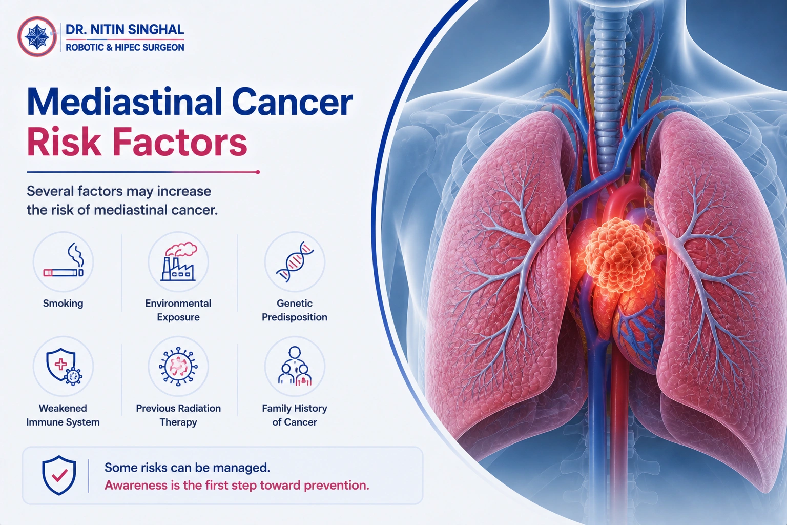
Mediastinal tumours are relatively uncommon and have a different risk profile depending on the tumour type. Having a risk factor does not mean a person will develop one.
Myasthenia Gravis
Around a third of patients with thymoma have this associated autoimmune condition.
Age
Thymic tumours are more common in adults aged 40–60; germ cell tumours often affect younger adults.
Prior Radiation Exposure
Previous radiation to the chest is linked to a higher risk of certain mediastinal tumours.
Underlying Lymphoma
Lymphomas can present as a mediastinal mass, particularly in younger patients.
Genetic Syndromes
Certain inherited conditions are associated with a higher risk of germ cell or neurogenic tumours.
Autoimmune Conditions
Some autoimmune disorders are more frequently seen alongside thymic tumours.
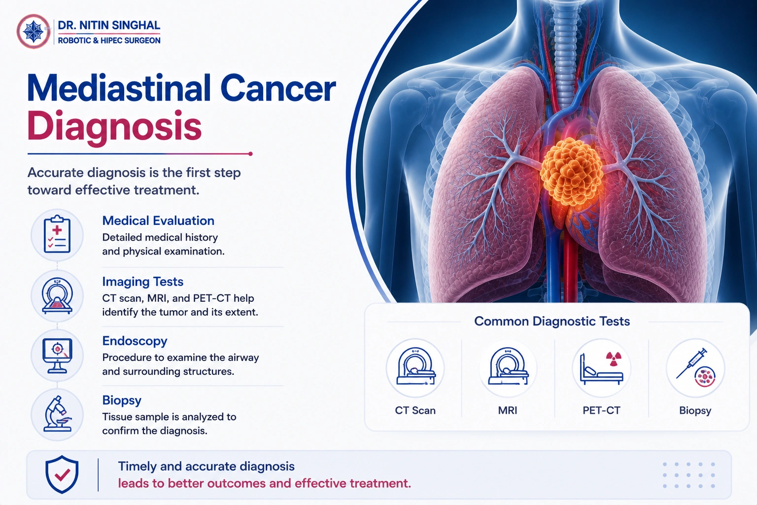
How Is Mediastinal Cancer Diagnosed?
Clinical evaluation — a review of symptoms, medical history and a physical examination.
Imaging (CT / MRI chest) — cross-sectional scans to locate the tumour and assess its relationship to the heart and great vessels.
Biopsy — a CT-guided needle biopsy or mediastinoscopy to obtain tissue for diagnosis.
Blood tumour markers — including AFP and beta-hCG, used when a germ cell tumour is suspected.
PET scan — used selectively to assess disease extent, particularly for lymphoma or suspected malignancy.
Pulmonary function tests — to assess lung function and fitness ahead of planning surgery.

How Mediastinal Cancer Is Treated
Treatment is planned individually and depends heavily on tumour type, decided by a multidisciplinary team.
Surgery
Removal of the tumour, often the primary treatment for thymomas and localised, resectable masses.
Chemotherapy
Used as primary treatment for lymphomas and some germ cell tumours, or alongside surgery for others.
Radiation Therapy
Considered after surgery in selected thymic tumours, or as primary treatment for some lymphomas.
Targeted / Immunotherapy
Considered for selected patients based on tumour biology and molecular testing.
Multidisciplinary Planning
Surgical, medical and radiation oncology teams jointly plan the treatment sequence.
Follow-Up Surveillance
Structured monitoring after treatment to check for recurrence.
Why Choose Dr. Nitin Singhal?
Robotic & HIPEC Cancer Surgeon
Focused practice in robotic-assisted and complex cancer surgery, including thoracic oncology.
Mediastinal & Thoracic Specialisation
Regular experience with mediastinal, lung and other thoracic cancers.
Training & Qualifications
MBBS, MS (General Surgery), MCh (Surgical Oncology) with training at Tata Memorial Centre, Mumbai.
18+ Years of Experience
Extensive experience across robotic and complex cancer surgery.
Hospital Affiliations
Operates at established hospitals in Ahmedabad with the infrastructure major thoracic surgery requires.
Multidisciplinary Cancer Care
Coordinated planning with medical oncology, radiation oncology and pathology colleagues as needed.

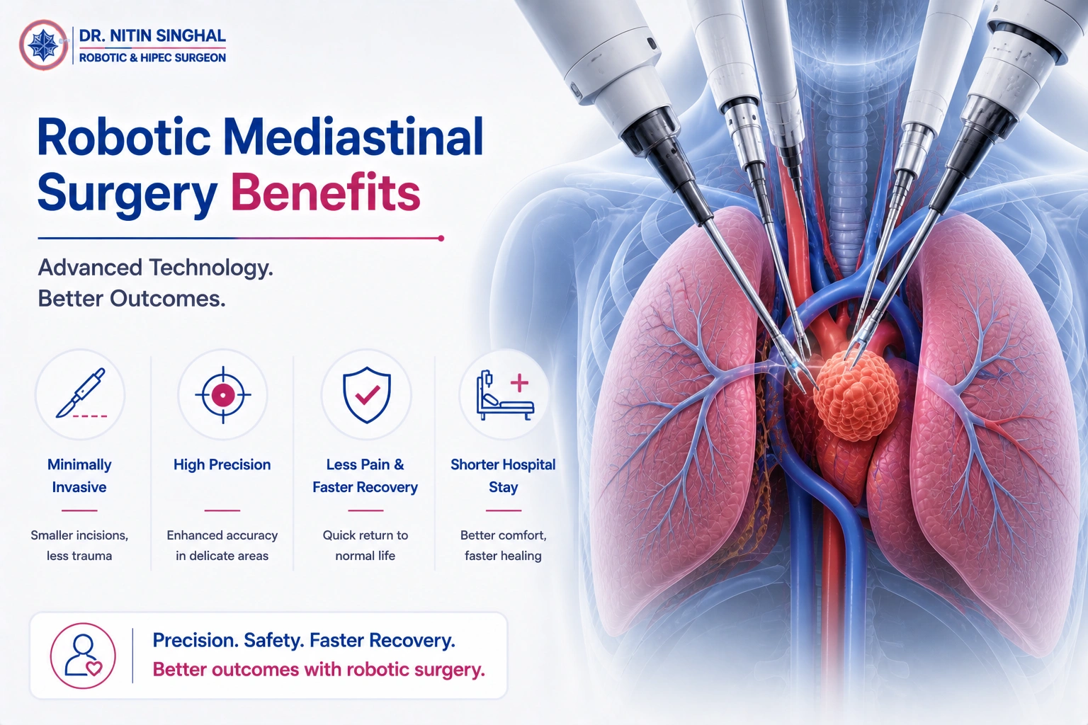
What Is Robotic Mediastinal Cancer Surgery?
Robotic-assisted surgery uses a surgical system with wristed instruments and a magnified 3D camera, controlled entirely by the surgeon from a console beside the patient. The robot does not operate independently — every movement is directed by the surgeon in real time.
The procedure is performed through a few small incisions in the chest wall rather than a full sternotomy (splitting the breastbone) or large thoracotomy. The magnified visualisation can support the meticulous dissection needed in the confined space of the mediastinum, close to the heart and great vessels — one of the most technically demanding areas of thoracic surgery. This differs from conventional open surgery, which requires a larger incision and direct visualisation by the surgeon.
Why Robotic Surgery May Be Considered
These are potential, patient-specific advantages in suitable cases — not guarantees, as outcomes depend on the individual patient and procedure.
Smaller Incisions
Several small incisions rather than a full sternotomy, in suitable cases.
Less Post-Operative Pain
Avoiding the breastbone incision may be associated with reduced chest wall pain during recovery.
Reduced Blood Loss
In appropriate cases, robotic technique can help limit intraoperative bleeding.
Earlier Mobilisation
Some patients are able to move around sooner after minimally invasive surgery.
Shorter Hospital Stay
In suitable patients, hospital stay may be shorter than after open surgery.
Precise Dissection Near the Heart
Magnified 3D vision can assist precision when working close to the heart and great vessels.
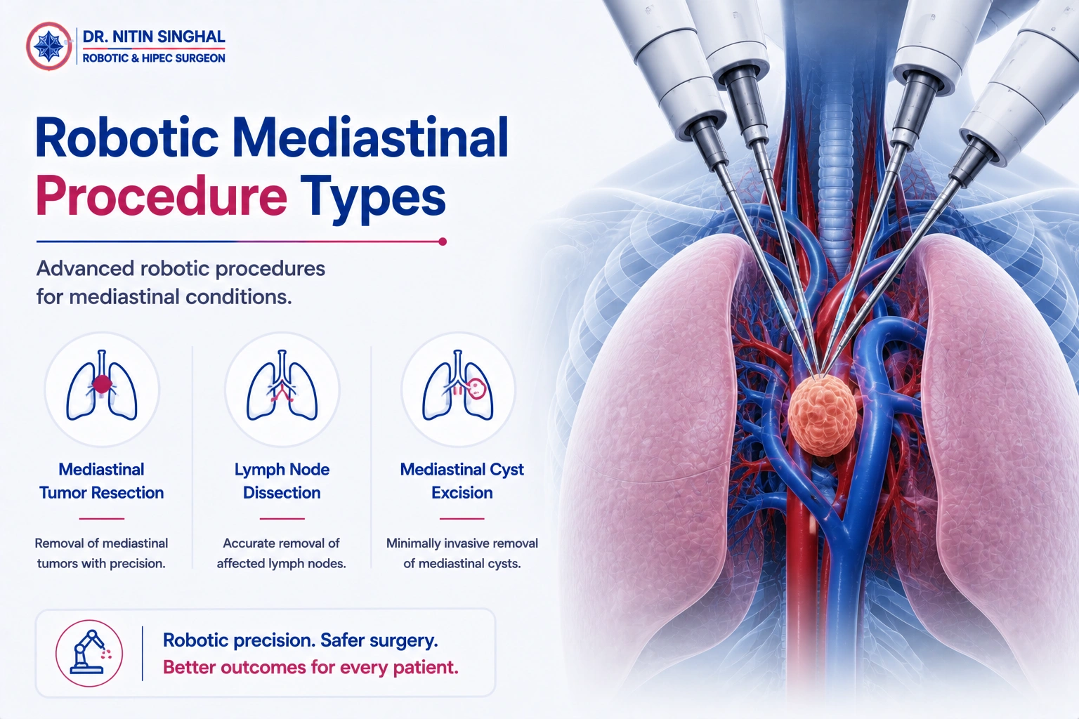
Types of Robotic Mediastinal Procedures
The specific procedure recommended depends on the tumour's location within the mediastinum and its type.
Robotic vs Laparoscopic vs Open Surgery
A general comparison of approach — actual outcomes for any specific patient depend on the procedure and case.
| Feature | Open Surgery | Laparoscopic Surgery | Robotic Surgery |
|---|---|---|---|
| Incisions | Larger (sternotomy / thoracotomy) | Smaller | Smaller |
| Visualisation | Direct | Camera-assisted, 2D | Enhanced 3D visualisation |
| Instrument Movement | Conventional, surgeon's hand | Limited range of motion | Wristed, surgeon-controlled robotic instruments |
| Recovery | Procedure-dependent | Procedure-dependent | Procedure-dependent |
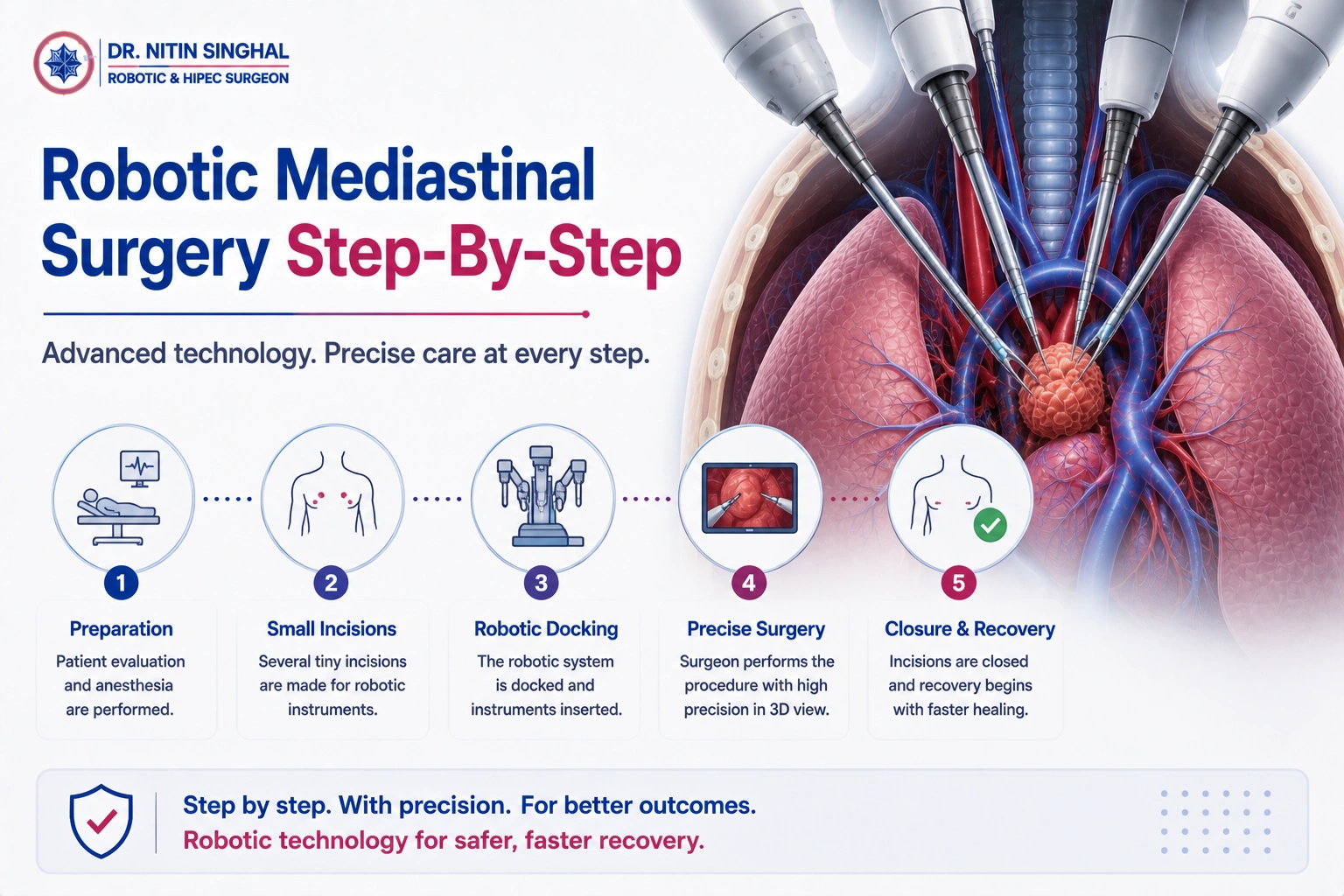
How the Surgery Works, Step by Step
Pre-operative evaluation — final review of imaging, staging and overall fitness for surgery, including lung function.
Anaesthesia — the patient is placed under general anaesthesia, typically with single-lung ventilation.
Small chest incisions — a few small incisions are made to introduce the robotic ports.
Robotic system positioned — the robotic arms are docked and the surgeon operates from the console.
Tumour and structures assessed — the mass and its relationship to the heart, great vessels and nerves are carefully evaluated.
Tumour removed — the thymus or mediastinal mass is dissected free and removed, preserving surrounding structures where possible.
Lymph nodes assessed — nearby lymph nodes are sampled where appropriate, for accurate staging.
Chest drain placed — a temporary chest tube is placed to drain fluid and air as the lung re-expands.
Incisions closed — the small incisions are closed, and the patient is moved to recovery.

Before Surgery & Recovery Afterwards
Investigations & Review
Final blood tests, imaging review and a medication review before the procedure.
Lung Function Assessment
Pulmonary function testing to confirm fitness for single-lung ventilation during surgery.
Anaesthesia Assessment
A pre-anaesthesia check to confirm fitness for the procedure.
Chest Drain & Pain Management
Structured post-operative care, including chest tube management and pain control.
Breathing Exercises
Guided breathing exercises to support lung re-expansion and reduce complications.
Return to Activity & Follow-Up
A structured plan for returning to normal activity, with scheduled follow-up visits.
Risks & Possible Complications
As with any major surgery, mediastinal surgery carries risks. These should be discussed in detail with your surgical team before deciding on treatment.
Bleeding
As with any surgery, there is a risk of bleeding during or after the procedure, particularly given proximity to the heart and great vessels.
Infection
Wound or chest infection is a possible complication of any thoracic surgery.
Nerve Injury
A risk to the phrenic or recurrent laryngeal nerve, which can affect breathing or voice.
Air Leak
Temporary leakage of air from the lung, usually managed with the chest drain.
Conversion to Open Surgery
In some cases, the procedure may need to be converted to open surgery for safety.
Myasthenic Crisis
A rare but serious risk in thymectomy patients with myasthenia gravis, requiring close monitoring.
Myths vs Facts
What Our Patients Say
Live reviews collected from Google, reflecting real patient experiences with Dr. Nitin Singhal and his team.
Cancer Care, Explained on Video
Patient-friendly explainers on cancer surgery, diagnosis, recovery and advanced robotic treatment from our channel.
Common Questions
What is robotic mediastinal cancer surgery?
Is robotic surgery suitable for every mediastinal tumour?
What is a thymectomy?
What is the recovery time after robotic mediastinal surgery?
How long will I stay in the hospital?
Is robotic surgery better than open surgery?
Will I need further treatment after surgery?
What happens to the removed tissue?
What are the risks of robotic mediastinal surgery?
How do I know whether I am a candidate?
Considering Robotic Mediastinal Cancer Surgery?
Get an expert evaluation to understand your diagnosis, treatment options, and whether robotic surgery may be appropriate for you.